
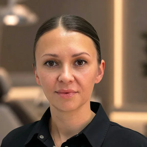
Anna Oleksandrivna
Chief dentist, general dentist
6 years of experience · KMU UANM
A digital X-ray of one to three teeth that the dentist takes during your visit. It shows the condition of the tooth, its root canals and the tissue around the root.
Reviewed by a dentist
Anna Oleksandrivna
Chief dentist, general dentist
Updated:
What we treat and how: pick a step
Sensor
A small digital sensor is placed in your mouth next to the tooth in question.
You wear a protective apron, and the X-ray tube takes the image in a fraction of a second.
The image appears on screen right away: it shows 1–3 teeth, their roots, canals and the bone around the root.
On the X-ray, the dentist finds hidden cavities: between the teeth and under old fillings.
During root canal treatment, the X-ray shows whether the instrument has reached the full length of the root and whether the canal is tightly filled.
Digital sensor
An electronic sensor instead of X-ray film.
Tooth
The X-ray shows the condition of the tooth and its root canals.
Bone
The X-ray also shows the tissue around the root.
X-ray tube
Takes the image in a fraction of a second.
Sensor
Captures the image. One X-ray on a digital sensor delivers a dose of 1–3 µSv.
Teeth
A periapical X-ray shows 1–3 teeth in fine detail.
Roots and canals
The X-ray shows the fine structures of the root.
Bone
The dentist assesses the bone around the root.
Cavity between teeth
Decay on the contact surfaces of the teeth shows up clearly on the X-ray.
Cavity under a filling
The X-ray reveals decay under an old filling.
Filled canal
The X-ray shows whether the canal is tightly filled.
Root tip
The dentist checks whether the instrument has reached the full length of the root.
A periapical dental X-ray is a digital X-ray of 1–3 teeth taken on a digital sensor during your visit, so the dentist can assess the tooth, its root canals and the bone around the root. It is only taken as part of a consultation or treatment and costs 200 UAH.
Price
200 UAH
per price list, as part of a consultationWhat it shows
1–3 teeth
roots, canals, boneSteps
5 steps
protective apron, sensor, X-ray, image, review by the dentistResults
In 2 seconds
the image is on screen during your visitAn electronic sensor instead of X-ray film. The radiation dose is 80–90% lower than on film-based machines.
The X-ray shows whether the instrument has reached the full length of the root and whether the canal is tightly filled.
During the X-ray you wear a special lead apron.
The X-ray is stored in your electronic patient file and can be sent by email or messenger.
The X-ray is taken only during a consultation with a dentist, and the image appears on screen within seconds.
You put on a special protective apron.
A small digital sensor is placed in your mouth next to the tooth in question.
The X-ray tube takes the image in a fraction of a second.
The X-ray appears on the computer screen right away.
The dentist reviews the X-ray and saves it to your electronic patient file.
Prices are per the price list. The dentist draws up a treatment plan with costs after an exam and diagnostics.
No. A periapical X-ray is only taken during a consultation with a dentist. It has to be interpreted by a dentist and is part of the diagnostics, not a stand-alone service.
To find cavities between the teeth and under old fillings, to assess a tooth when pulpitis or periodontitis is suspected, and to check root canal treatment. Also to assess the bone around a root or an implant, for injuries, root fractures and cracks, and before and after oral surgery or prosthetic treatment.
One periapical X-ray on a digital sensor delivers 1–3 µSv. The annual limit allowed for preventive medical exams is 1,000 µSv.
If the treatment requires it, several X-rays are taken in one visit, for example before and after the canals are filled.
When medically necessary, a periapical X-ray can be taken in the second trimester with a special protective apron. The dentist makes the decision after weighing the benefits and risks.
Yes. The digital X-ray is stored in our database. On request, we will send it to your email or messenger or save it to a drive.
A periapical X-ray shows 1–3 teeth in fine detail, including the small structures of the root. A panoramic X-ray shows the full set of teeth in both jaws and the jaw joints, but individual canals are harder to see on it.
All dentists
Chief dentist, general dentist
6 years of experience · KMU UANM

Chief dentist, oral surgeon, prosthodontist
9 years of experience · UANM

General dentist
7 years of experience · DSMU

Orthodontist
12 years of experience · DNMU

Oral surgeon, prosthodontist
8 years of experience · UANM

General and pediatric dentist
13 years of experience · Bogomolets NMU

General dentist
16 years of experience · LNMU

Oral surgeon
8 years of experience · UMSA

General dentist, prosthodontist
5 years of experience · KhNMU

General dentist
9 years of experience · KMU UANM

General dentist
8 years of experience · VNMU

General dentist
5 years of experience · LNMU
Endodontist
15 years of experience · DonNMU
I recommend it 100%. Professional, high-quality and painless. The office has an X-ray machine, so it's convenient to take an image 'on the spot' for further treatment.
I had a CT scan at this dental office and was very pleased! The staff are friendly, attentive and professional. The atmosphere is cozy, which helps you feel calm during the procedure. The service is excellent, everything is well organized and comfortable. I recommend it!😌🙂👍🏻
I want to thank the dental practice's administrator for her professionalism, friendliness and excellent organization! 😊 She really helped me choose the right X-ray, explained everything in detail and suggested the best option. It's nice when patients are treated with attention and care. Thank you for the great service!
I had a CT scan, everything went well and fairly quickly. The administrator is very friendly. You can see they're very busy, so book in advance ❤️
They did my CT scan very quickly. Customer focus at the highest level: despite the unpredictable power outages, they called me and scheduled the appointment. I recommend them!
Quick, high-quality CT scan. The staff's explanations are polite and to the point, without small talk or fake smiles
I was at my first visit with Maryna Ihorivna, a very professional doctor. She showed me photos of my problem teeth, gave me clear advice and told me what needs to be worked on. They also did a CT scan, on which the doctor pointed everything out to me in more detail. Big thanks to the doctor's assistant too!
I get panoramic X-rays done for my whole family here: convenient, no queues. The result is instant and informative. I recommend it: nice administrators and a convenient location near the metro. My dental office is on L. Lukianenka St
I'm very grateful to Eurozet for their speed. They booked me an appointment literally an hour after I contacted them. They did a CT scan, clearly explained the situation at that moment and made a further treatment plan. The staff are very friendly. Special thanks to the young woman at reception.
Everything is great! I had a CT scan and got all my answers at the consultation, thank you!
I had a CT scan at this dental office and have only positive impressions. Pleasant staff, cleanliness, attention to the patient and efficient work. You can feel the professional approach and care for people. They do everything well and fast!
I came to this dental office for a CT scan and a consultation to be offered different options for solving my problem. I was left with positive feelings and plan to have my treatment here.
I booked a CT scan the same day; the operator helped me pick a convenient time, the staff are very friendly, and I got the CT within 10 minutes Very pleased, I'll definitely come again
The Eurozet website will close and another website will open.
Go to Google reviewsAny questions? Call us: +38 (098) 661 10 99
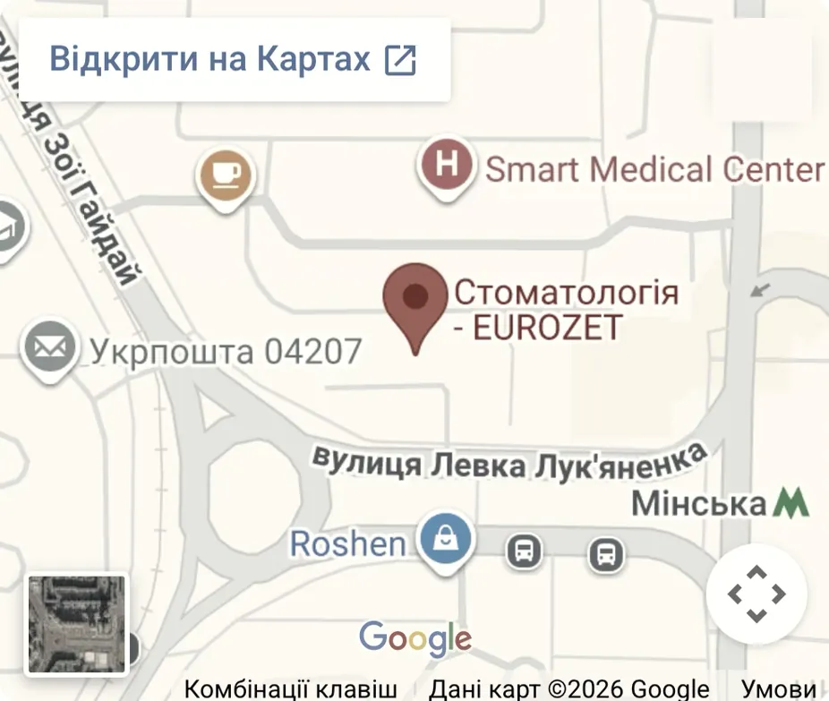
Minska metro station
21 Levka Lukianenka St
Every day, 9:00 to 21:00
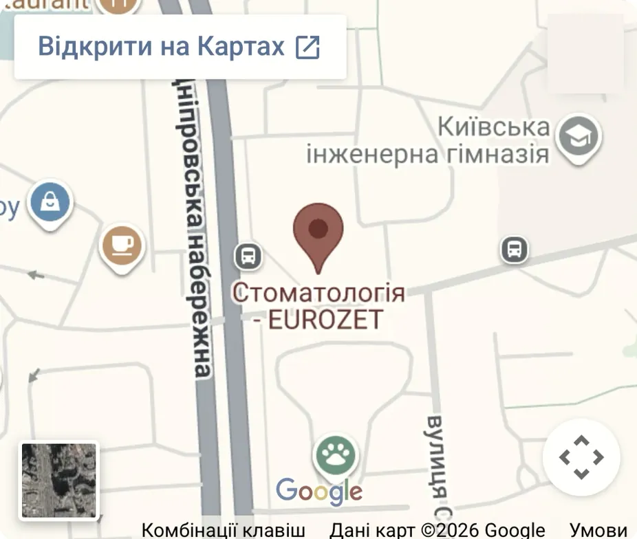
Osokorky metro station
14B Kniazhyi Zaton St
Every day, 9:00 to 21:00
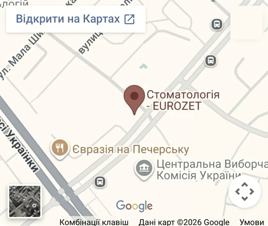
Pecherska metro station
12 Henerala Almazova St
Every day, 9:00 to 21:00













A digital X-ray system uses a sensitive electronic sensor instead of X-ray film. As a result, the radiation dose is several times lower, and the image appears on the monitor within seconds. The dentist can zoom in on the image on screen and examine small changes in the tooth and around the root. The X-ray is saved to your electronic patient file right away, and we can send it to you on request.
During root canal treatment, the dentist has to make sure the instrument has reached the full length of the root. After filling, it is important that the material fills the canal tightly, with no voids. The only way to see this is on an X-ray. The dose on a digital sensor is minimal, so when needed the dentist takes several follow-up images in one visit. Read more about the procedure itself on the root canal treatment page.
Per the price list, a periapical X-ray costs 200 UAH. It is not taken separately from a visit: the X-ray has to be interpreted by a dentist, so it is part of a consultation or treatment that has already been prescribed. A consultation with a treatment plan costs 500 UAH. If an X-ray is needed during treatment, the dentist takes it right there during the visit, with no separate appointment. The full diagnostics price list is on the Prices page.
Near Minska metro station is the Eurozet location in Obolon: 21 Levka Lukianenka St. The second location is in Pozniaky, 14B Kniazhyi Zaton St, next to Osokorky metro station. The third is in Pechersk, 12 Henerala Almazova St, next to Pecherska metro station. At all three locations, X-rays are taken right during your visit, every day from 9:00 to 21:00. Treatment that includes X-rays can be paid in installments through PrivatBank.
Thank you!
We will call you shortly at .
This window will close automatically


